Utilization of Bioanalytical Techniques in the Crime Scene
Introduction
Forensic evidence has become central to the criminal justice system for establishing the guilt or innocence of potential suspects, tracing the origins of illegal trade and determining whether or not crimes are related.
Such evidence can increase the efficiency of a criminal investigation by narrowing down the search for suspects, which in turn reduces the time needed to bring the perpetrator to justice. It can also help ensure successful prosecutions in court since links to a crime can be scientifically proven.
Forensic evidence includes a range of information or objects that are admissible in court. It can take many forms, including impression evidence, such as footprints and tool marks, trace evidence, such as fibres or chemicals, body fluids and DNA, drugs, firearms and explosives. Analysis of such evidence found at the crime scene can be used to both facilitate the investigation and secure the prosecution of guilty parties in court.
The nature of the forensic analysis varies according to the type of crime. It may be used to trace the origins of an illicit substance, identify persons who were present at the crime scene or reconstruct the series of events leading up to a criminal act.
Detection of traces of explosives, for example in improvised devices intended to cause mass devastation, can help defeat planned acts of terrorism. These technologies can also help inform the armed forces active in conflict zones.
Since there is such a broad scope of formats of potential evidence, it follows that numerous techniques must be employed in forensic analyses. Consequently, forensic science encompasses the application of knowledge from across all the sciences, including physics, chemistry, biology, computer science and engineering. A wide range of methodologies are thus employed by forensic scientists. These include various forms of analytical and imaging techniques, such as spectroscopy and spectral sensing.
Analyses may need to determine whether there are chemicals in the blood of a victim and if so, from where they originated. Comparisons of DNA, found in the form of hair or body fluids at the crime scene, and that of potential suspects can help identify whether they are implicated in the crime. Similarly, fibers from clothing or paint found at a crime scene can be matched to items found in the possession of a suspect and help establish whether or not they were present at the crime scene. Analysis of gunpowder residues, spent bullets and cartridges can help establish the weapon that was used in a crime, from where the shot was made and even who fired it.
Therefore overall bioanalysis is a fundamental tool in forensic science, and continuing technological advances are achieving ever-greater capabilities, in terms of sensitivity, specificity, accuracy, efficiency, throughput, or data quality. In order to ensure that justice is served with the greatest confidence, it is critical that innovation in analytical methodologies are efficiently incorporated into forensic laboratories in a timely manner.
The National Institute of Justice (NIJ) is dedicated to improving the administration of justice through advancing scientific research. NIJ funds research and development to improve how law enforcement gathers and uses evidence. It also supports the enhancement and creation of tools and techniques to identify, collect, analyze, interpret and preserve evidence.
Last year alone NIJ funded 50 projects researching developments in forensic science for criminal justice purposes. These include a range of studies into novel spectroscopic techniques, improved analysis of finger prints, identification of body fluids, and addition of entries in the national powder database, which was designed to help identify and match gunpowder found at crime scenes.
The latest analytical technologies developed with increased sensitivity, including laser-induced breakdown spectroscopy, deep ultraviolet spectroscopy, hyperspectral imaging, and ultraviolet Raman spectroscopy, are already finding applications in forensic science.
The greater capabilities provided by these techniques further increase the information that can be discerned from evidence obtained at crime scenes. This in turn gives the justice system more power to identify and prosecute perpetrators of crime.
Furthermore, the companies providing the instrumentation needed by forensic laboratories to conduct the analyses on samples they receive are continually updating and modifying the equipment they produce, based on the findings of the latest research, in order to enhance the analytical capabilities on offer.
This article introduces the latest bioanalytical and imaging techniques and equipment, which will be presented in more detail at Pittcon 2018, and describes how they can be applied in forensic analyses.
References
- Dutton GJ, et al. Cultivating the Next Generation of Forensic Scientists Through Science, Technology, Engineering, and Mathematics (STEM). J Forensic Res 2017, 8:4. Available at https://www.omicsonline.org/open-access/cultivating-the-next-generation-of-forensic-scientists-through-sciencetechnology-engineering-and-mathematics-stem-2157-7145-1000384.pdf
- National Institute of Justice. Forensics. https://www.nij.gov/topics/forensics/Pages/welcome.aspx
Chapter 1 – Using bioanalytical methods for the forensic study of evidence
The development of new technology has allowed for the evaluation of materials and substances of relevance to a legal inquiry beyond what the naked eye can see. Investigations now employ the laboratory for the analysis of a diverse range of evidence, and novel techniques are continuously being established.
For each new scientific development, it is important that there be a standardized methodology for implementation so that evidence is admissible in a court of law. Bioanalytical methods involve the quantitative measurement and analysis of biotics such as DNA and metabolites. The process begins with the forensic study of evidence such as blood, saliva, hair and skin cells which contain biomolecules that can be evaluated for links between the sample and the crime scene.
Pittcon 2018 will provide examples of the latest innovations in bioanalytical methods for forensics. This will include talks supplying details of novel methodologies for analyzing evidence, along with exhibits of current bioanalytical technology. The sessions will combine examples of new technologies that allow for more effective and efficient forensic investigations with the robust science that ensures excellent results.
Rapid DNA re-identification
Fingerprinting is a well-known form of forensic evidence but modern forensic approaches allow so much more. There are now numerous forms of biological evidence. In particular, DNA as a biometric identifier has revolutionized forensic science; current re-identification schemes however suffer from high latency and low portability.
At Pittcon 2018, Sophie Zaaijer’s talk ‘Democritizing DNA Fingerprinting’ will note the importance of re-identifying DNA and describe the development of ‘MinION sketching’- a method of DNA re-identification through the use of Single Nucleotide Polymorphisms (SNPs) that is portable, rapid, robust and inexpensive.
Using Oxford Nanopores MinION sequencer, the procedure of ‘MinION sketching’ does not require PCR, but instead analyzes low coverage shot-gun sequencing data and then compares the variants to a reference database of SNPs. MinION sketching can be applied to border control through the prevention of human trafficking, on-site crime scene re-identification of DNA samples and the rapid identification of victims after a mass disaster.
DNA sequencers for forensic applications
Additionally, Pittcon 2018 exhibitor ThermoFisher provide a number of products that yield integrated human identification solutions. They include instruments for real-time human identification through PCR, developed to allow forensic labs to achieve accuracy in a shorter time.
ThermoFisher’s 3500 Series Genetic Analyzers are the first class of analyzer instrument to feature a workflow specifically designed for human identification applications. The instrument is based on proven ThermoFisher Applied Biosystems capillary electrophoresis technology with the additional ability to provide step-by-step procedural control of data collection and real-time quality assessment.
Eppendorf also manufacture products with infallible results that adhere to the high level of regulatory requirements for utilizing DNA as forensic evidence. This includes precision instruments designed for processes starting with sample preparation through to the amplification and detection stage of forensic DNA profiling.
The Eppendorf PCR Thermocyclers offer the essential ability to obtain reproducible results quickly. During PCR, evaporation can cause the increased concentrations of components such as primers, leading to non-specific binding and a continuously changing concentration variable. The Eppendorf Mastercycler Pro employs the vapo.protectTM concept which reduces evaporation for consistent sample concentrations.
The analysis of glass as forensic evidence
It is also important to consider the implementation of objects, surfaces and materials as evidence for criminal investigations. This includes the analysis of glass as a contact trace material. When a pane of glass is broken, the minute glass fragments can be transferred to the person responsible.
Furthermore, distinctive variation in glass manufacturing allows for the discrimination of this trace evidence.
Jose Almirall will give a talk at Pittcon 2018 entitled ‘The Development of a LIBS Database for the Forensic Interpretation of Glass Evidence’, which will provide an overview of the use of Laser Induced Breakdown Spectroscopy (LIBS) for standardizing glass evidence analysis.
Previous research has found that Laser Ablation-Inductively Coupled Plasma-Mass Spectrometry (LA-ICP-MS) and the associated database of glass profiles provide a low classification error rate. At Pittcon 2018, Applied Spectra will be exhibiting analytical instruments based on laser ablation technology.
Their products supply rapid elemental and isotopic analysis without the need for large amounts of sample preparation. Applied Spectra’s J200 Femtosecond Laser Ablation Instrument is a custom-built tool allowing for efficient forensic analysis of materials through the tailoring of application requirements.
Alternately, LIBS can provide an inexpensive methodology with reduced complexity for glass analysis. The technique works on the principle that all elements emit light at frequencies that can be characterized when excited by a laser. The resulting chemical characterization of the glass sample can be used to prove an association between the sample and fragments collected at the crime scene.
Experiments comparing LIBS with other techniques for glass analysis, such as LA-ICP-MS, have found that LIBS provides comparable discrimination power. For forensic purposes, LIBS analysis requires a database of glass samples that can be used for comparison and the calculation of likelihood ratio estimates.
Exhibitors at Pittcon 2018 with LIBS equipment for glass analysis include Oxford Instruments and LTB Lasertechnik Berlin. Additionally, it will also be possible to speak to B&W Tek at Pittcon 2018, about their handheld LIBS analyzer – The NanoLIBS-Q.
The Vulcan handheld LIBS analyzers produced by Oxford Instruments provide rapid analysis in a second. The simple point-and-shoot system interface reduces user-related error whilst increasing the number of analyses that can be performed per day.
During forensic investigations it is important that samples are retained for the future and the Vulcan LIBS analyzer ensures the integrity of the sample by requiring less than one billionth of a gram to be eliminated during the process.
The Libspector from LTB Lasertechnik Berlin is suitable for performing LIBS analysis on solid, liquid and gaseous samples. It is available for forensic applications as a compact bench-top sample chamber with in-built laser protection. The safety lock and laser protection window permit safe observations without the need for additional precautions to be installed.
References
- O’ Shaughnessy, PE. Introduction to Forensic Science, Dental Clinics of North America 2001, 45; doi: https://www.ncbi.nlm.nih.gov/pubmed/11370451
- Zaaijer, S, Gordon, A, Speyer, D. et al. Rapid DNA Re-Identification for Cell Line Authentication and Forensics, bioRxiv 2017; doi: https://doi.org/10.1101/132381
- Thermo Fisher: Integrated Human Identification Solutions. http://www.thermofisher.com/uk/en/home/industrial/forensics/human-identification.html
- Eppendorf: Forensic DNA typing. https://www.eppendorf.com/PT-en/applications/forensic/forensic-dna-typing/
- Forensic Access: Glass Analysis. http://www.forensic-access.co.uk/glass-trace-evidence-forensic/
- Naes, BE, Umpierrez, S, Ryland, S. et al. A comparison of laser ablation inductively coupled plasma mass spectrometry, micro X-ray fluorescence spectroscopy, and laser induced breakdown spectroscopy for the discrimination of automotive glass, Spectrochimica Acta Part B 2008, 63, doi: https://doi.org/10.1016/j.sab.2008.07.005
- Cahoon, EM and Almirall, JR. Wavelength dependence on the forensic analysis of glass by nanosecond 266 nm and 1064 nm laser induced breakdown spectroscopy, Applied Optics 2010, 49, doi: https://doi.org/10.1364/AO.49.000C49
- Barnett, C, Cahoon, EM and Almirall, JR. Wavelength dependence on the elemental analysis of glass by Laser Induced Breakdown Spectroscopy, Spectrochimica Acta Part B 2008, 63, doi: https://doi.org/10.1016/j.sab.2008.07.002
- Applied Spectra J200 Femtosecond Laser Ablation Instrument. https://appliedspectra.com/products/j200-femtosecond-la.html
- Oxford Instruments Hitachi Handheld LIBS Analysers: Vulcan Range. https://hha.hitachi-hightech.com/en/product-range/products/handheld-xrf-libs-analysers/handheld-libs-analysers
- LTB Lasertechnik Berlin: Libspector. http://www.ltb-berlin.de/en/products/accessories/libspector/
Pittcon Tracks
Biological molecules and xenobiotics (drugs, toxins) and their metabolites; study of biological systems; biosensors; forensic science and toxicology
Identification, quantitative measurement, extraction, and quality assurance of cannabis-based and psychedelic products
Environmental detection and monitoring; energy production and storage; sustainability, climate, and green chemistry; food science/safety and agriculture
Instrumentation, detection, and sensors; laboratory information systems, data analysis, and artificial intelligence; characterization and processing of nanomaterials; art and archeology
Evaluating chemical composition and properties/activities of medicinal drugs and biologics; high-throughput screening and process control; drug discovery and design; personal care and consumer products
Leadership and power/soft skills; career navigation, DEI (diversity, equity and inclusion), communication, and entrepreneurship; education and teaching and more
Chapter 2 – Emerging Technologies for Trace Evidence Analysis
Forensic evidence admissible in court plays a vital role in the criminal justice system. Analysis of materials obtained from a crime scene or the clothing of potential suspects can provide a picture of events surrounding a crime and help determine the guilt or innocence of defendants.
The National Institute of Justice (NIJ) is the research, development and evaluation agency of the US Department of Justice. It is dedicated to improving knowledge and understanding of crime and justice issues through science. NIJ’s mission is to advance scientific research, development, and evaluation to enhance the administration of justice and public safety.
During a crime, tiny fragments, such as hairs, fibers, paint and fragments of glass can be transferred between individuals and objects. These are referred to as trace evidence and can provide important information about what happened at the time of the criminal activity and who was present. For example, a hair found on the clothing of a suspect can confirm contact with the victim.
Careful collection and analysis of materials from a crime scene can yield a wealth of information about the origin of a sample. Scientists examine the physical, optical and chemical properties of trace evidence and use a variety of tools to find and compare samples, and look for common origins across items.
There is an ongoing need for these analyses to be performed on ever smaller or lower quality samples without sacrificing the validity of the results. NIJ’s continuing investment into the research and development of analytical chemistry and bioanalytical science helps ensure that objective, rigorously tested methodologies are developed to enable reliable analysis of the most challenging samples.
Many advances in mass spectrometry, electrophoresis, applied spectroscopies, microscopy and microfluidics have shown successful application to forensics, and many more show great potential.
At Pittcon 2018, Gregory Dutton of NIJ will be providing an overview of NIJ’s research and development portfolio, presenting details of projects supported in the past and highlighting new funding opportunities, in his presentation entitled ‘Forensic Science Research and Development Funding Program at the National Institute of Justice: Opportunities in Analytical Chemistry, Applied Spectroscopy and Bioanalysis’.
Since forensic analyses require the utilization of specialist techniques from a broad range of disciplines, it would be unfeasible for research laboratories to undertake research and development across all areas of science and engineering they need to employ in order to address the wide variety of tests required to solve crimes and bring the perpetrators to justice.
The NIJ’s long-term strategy to undertake research to advance forensic science and train scientists of the future allows the required innovation without distracting forensics laboratories from conducting the batteries of tests needed for justice to prevail.
Research success stories include the development of multistage mass spectroscopy that allows identification, e.g., of drugs in blood or urine, to be made more rapidly without the need for derivatization steps or the use of hazardous solvents. Similarly, the validity of examination of trace evidence materials has been improved by elemental analysis methods.
Microbiome analysis
Each individual is home to around 100 trillion microbial cells, on the skin, and in the eyes. mouth and gut, which comprise their microbiome. A microbiome can include more than 10,000 different microbial species, the precise combination of which is determined by the environments visited and the foods eaten. Consequently, the composition of microbial organisms associated with skin is unique to an individual because each individual has a unique range of experiences.
NIJ have been funding microbiome research for many years, but more recently research has included the investigation of forensic applications of microbiomes.
These are categorized into three main areas: the necrobiome — the community of organisms found on or around decomposing remains — which provides an indication of the time-since-death; soil microbiome, which can link a victim, suspect, or evidence to a particular outdoor environment; and the trace human microbiome — microbes on our skin and the surfaces and objects we interact with —which provides the potential for associating people with evidence and environments.
A unique study assessing whether a person’s traits and lifestyle can be predicted from their skin microbiome will be described at Pittcon 2018 by Jack Gilbert of the University of Chicago in a presentation entitled ‘The Burglary Microbiome Project: Detecting Personal Microbiome Signatures at Artificial Crime Scenes’.
Methodologies used in analysis of trace evidence
Since trace evidence can comprise virtually any substance, a broad scope of very different methodologies is used by forensic scientists to solve crimes. These are based on knowledge from all branches of sciences, including biology, chemistry, physics and mathematics, and enabled by a range of sophisticated technologies. It is this fusion of science and technology that provides forensic scientists with the power to unravel crime scenes.
There have been tremendous advances in technology in recent years. Forensic scientists now have capabilities that would have been considered pure fantasy only a matter of years ago.
Analysis of paint residue can determine the age and make of a car, gunshot particles can be distinguished from dust, dirt and other fibers, the presence and exact composition of drugs can be established, soil samples can be matched to a specific locality by determining the precise mineral and organic components – and the list continues to grow.
The technologies used to achieve these remarkable connections include spectroscopy, spectrophotometry, gas chromatography, microcrystalline testing, and X-ray diffraction.
Analysis of gunshot residue
Detection of gunshot residue can play an important role in determining where a crime took place and who pulled the trigger, for example, by establishing the presence of gunshot residue on the clothes of a suspect.
Traditionally, gunshot residue detection has relied on the presence of heavy metals, but with heavy metals becoming less common in ammunition alternative analyses were needed. Vibrational spectroscopy, such as Raman microscopy and Fourier transform infrared spectroscopy, has proved to be a particularly useful analytical tool in the detection and characterization of gunshot residue.
It requires minimal sample preparation, is nondestructive and detects both organic and inorganic constituents. Vibrational spectroscopy automatically scans large areas of the collection tape and produces a spectroscopic fingerprint detailing the various components of the gunshot residue.
A complex chemical reaction occurs when a firearm is discharged and the precise nature of this is determined by the exact chemical composition of the ammunition and the specific firearm used. The presence of gunshot residue discharged into the immediate surrounding during firing can be accurately determined using vibrational spectroscopy.
Furthermore, analysis of the gunshot residue using provides characteristic spectra that can provide information about the type of ammunition and weapon used.
In his presentation at Pittcon 2108, ‘Vibrational Spectroscopy and Advanced Statistics for Detection and Characterization of Gunshot Residue’ Igor Lednev of the University at Albany, will be describing how gunshot residues can have been successfully classified according to caliber based on the spectra obtained from vibrational spectroscopy.
Raman spectroscopy is often favored over infrared spectroscopy for chemical identification since it causes fewer peaks and is not swamped with water absorption lines so spectra are easier to interpret. It has the potential to detect a wealth of information from both solid and liquid samples, but the quality of information obtained is determined by the instrumentation used.
Raman spectroscopy is based on the scattering of high-intensity light. It is therefore important to collect as many photons as possible while rejecting scattered laser light efficiently.
Ocean Optics will be at Pittcon 2018 to discuss the capabilities of their high-sensitivity spectrometers, including the Maya2000 Pro.
Toolmark analysis
Forensic analysis is often required to ascertain whether a bullet or cartridge case found at the crime scene was fired from a particular weapon, e.g., one found in the possession of a suspect. This is achieved using toolmark analysis, which studies the specific marks made on the bullet or cartridge case by the weapon from which it was fired. The marks on a spent bullet are unique to the weapon and so microscopic examination of these marks allows firearm examiners to assess the likelihood of it having been expelled from a given firearm.
Traditionally, a two-dimensional representation of the bullet or cartridge surface, known as The Integrated Ballistics Identification System, has been used by forensic laboratories for toolmark analysis.
However, a range of three-dimensional (3D) scanning technologies is now available to facilitate toolmark analysis, such as TopMatch produced by Cadre Forensics. These provide a high-resolution 3D digital image of the surface topography, which directly correlates to the actual physical surface.
Examination of the digital image thus allows easy sharing of the evidence for simultaneous remote evaluation. Furthermore, the images can be automatically compared with a database and catalogued by case for future reference.
A specialized viewing software that allows annotation of the areas of similarity identified during toolmark analysis will be presented at Pittcon 2018 by Cadre Forensics in a session entitled ‘3D Surface Topography Analysis and Virtual Microscopy for Firearm Forensics’.
References
- Bueno J, et al. Raman Spectroscopic Analysis of Gunshot Residue Offering Great Potential for Caliber Differentiation. Anal Chem 2012;84(10):4334–4339.
- Bueno J and Lednev IK. Raman Microspectroscopic Chemical Mapping and Chemometric Classification for the Identification of Gunshot Residue on Adhesive Tape. Anal Bioanal Chem 2014;406:4595 4599.
- Bueno J and Lednev IK. Attenuated Total Reflectance-FT-IR Imaging for Rapid and Automated Detection of Gunshot Residue. Anal Chem 2014;86(7):3389–3396.
- Duez P, et al. Development and Validation of a Virtual Examination Tool for Firearm Forensics. J Forensic Sci. 2017; doi: 10.1111/1556-4029.13668. [Epub ahead of print]. Available at http://onlinelibrary.wiley.com/doi/10.1111/1556-4029.13668/full
- Dutton GJ, et al. Cultivating the Next Generation of Forensic Scientists Through Science, Technology, Engineering, and Mathematics (STEM). J Forensic Res 2017, 8:4. Available at https://www.omicsonline.org/open-access/cultivating-the-next-generation-of-forensic-scientists-through-sciencetechnology-engineering-and-mathematics-stem-2157-7145-1000384.pdf
- National Institute of Justice. Forensics. https://www.nij.gov/topics/forensics/Pages/welcome.aspx
- Xie F, et al. Automated bullet-identification system based on surface topography techniques. Wear 2009;266(5–6, 15):518 522.
Chapter 3 – Identification and Detection using Raman Spectroscopy
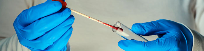
Body fluid analysis
The detection and identification of body fluids at a crime scene can provide essential information about who was present and the events occurring around the time of the crime. The presence of body fluids, their location and their DNA profile can significantly aid police investigations by presenting a picture of the circumstances of the crime.
The presence of body fluids at a crime scene, even if they have been wiped up or covered, can be detected using alternative light sources. Extraction and sequencing of DNA from any body fluids found at a crime scene, or on a suspect or victim, can be used for identification purposes.
However, alternative light sources will not typically allow for differentiation between different body fluids. Blood, sweat, saliva, faeces, urine, vaginal fluid and semen are all potential sources of evidence, depending on the nature of the crime.
Although blood is readily identified using color change tests, such as luminol or phenolphthalein, it is not so easy to identify the origin of other stains. Identification of urine, vaginal fluid and semen is particularly useful when ascertaining whether a sexual crime has been committed.
Historically, there was no single method for the analysis of all body fluids and identification was largely based on determining the presence of known components of a given body fluid, for example amylase in saliva, and urea in urine.
Fourier transform infrared spectroscopy and Raman spectroscopy now offer the potential to identify biological stains at a crime scene without detriment to the sample.
Each body fluid has its own characteristic spectral signature and these have been used to correctly classify stains, irrespective of whether they were fresh or dried and the color or nature of the material on which they were found. Furthermore, contamination with soaps, milk, juices, and lotions did not give rise to erroneous conclusions.
At Pittcon 2018, Igor Lednev of the University at Albany will be detailing the identification of traces of body fluids in his presentation ‘A Universal Method for Biological Stain Characterization Using Raman Spectroscopy: From Body Fluid Identification to Phenotype Profiling’.
It was also possible to distinguish animal blood from human blood and menstrual blood from peripheral blood. Furthermore, advanced statistical analysis of the spectroscopic data enabled discrimination between Caucasian and African American donors with over 80% accuracy.
Renishaw will be available at Pittcon 2018 to discuss the capabilities of their new inVia Qontor Raman microscope, which includes LiveTrack™ focus tracking technology that allows accurate analysis of samples with uneven, curved or rough surfaces.
Optimum focus is maintained without the need for time consuming manual focusing, pre-scanning or sample preparation. The highly efficient optical design provides the best Raman data from minute traces of material.
The inVia Qontor Raman microscope can be used with the Bruker Dimension Icon AFM, which provides high-performance surface characterization and the flexibility to perform nearly every atomic force microscopy measurement type at resolutions previously only obtained by extensively customized systems.
This additional pairing demonstrates the extreme flexibility of the Renishaw inVia confocal microscope, and its ability to interface to a wide range of instruments employing many analytical techniques.
Detection of explosives
In the current climate where there is a high risk of terrorist bombings and improvised explosive devices, the ability to detect traces of explosives from a distance is of increased importance.
Since there are many different explosives, which are often present in small quantities amidst numerous background components, it has proved challenging to develop an effective means of detection that provides the required selectivity and sensitivity.
Raman spectroscopy is well suited to detecting explosive traces remotely since explosives give distinct, narrow Raman spectral bands. The selectivity and sensitivity can be further enhanced by using a deep-ultraviolet (DUV) light source.
Although visible and infrared Raman spectroscopy wavelength provides deeper sample penetration, the DUV enhancement allows for higher signal levels from traces of explosives.
DUV resonance Raman spectroscopy is thus a promising potential candidate for stand-off detection of explosives. Furthermore, it is possible for the components of this technology to be produced in a compact, portable format suitable for transporting to various locations where screening is to be conducted.
A highly sensitive algorithm has also been developed for the detection of explosives under low signal-to-noise situations.
The development of prototype explosive detectors based on DUV resonance Raman spectroscopy will be presented by Balakishore Yellampalle at Pittcon 2018 in a presentation entitled ‘Compact Deep Ultraviolet Resonance Raman Explosive Detector’.
Ibsen Photonics, who will be attending Pittcon 2018, produce the FREEDOM™ HR-DUV compact deep UV spectrometer. It offers high performance whilst being compact enough to be portable and robust enough to operate in demanding environmental conditions.
Spectrometers in the FREEDOM range support many different detector systems so can be tailored to suit a specific application. Furthermore, they allow the use of existing electronics and software.
Horiba Scientific will also be at Pittcon 2018 presenting their SWIFT™ ultra-fast Raman imaging system. Real-time detailed Raman images detailed can now be acquired on second/minute timescales with integration times down to 1ms. Importantly, SWIFT retains the true confocal performance of the HORIBA Scientific Raman systems, ensuring optimized spatial resolution for effective analysis of thin layers.
References
- Sikirzhytski V, et al. Discriminant Analysis of Raman Spectra for Body Fluid Identification for Forensic Purposes. Sensors 2010;10(4):2869-2884.
- Yellampalle B, et al. Multiple-excitation-wavelength resonance Raman explosives detection. Proc. SPIE 8018, Chemical, Biological, Radiological, Nuclear, and Explosives (CBRNE) Sensing XII, 801819. June 03 2011. Available at http://www.wvhtf.org/wp-content/uploads/2015/08/MEWRRED-DSS-11.pdf
- Yellampalle B, et al. Dual-Excitation-Wavelength Resonance-Raman Explosives Detector. Proc. SPIE 8710, Chemical, Biological, Radiological, Nuclear, and Explosives (CBRNE) Sensing XIV, 87100Z. May 29 2013. Available at http://wvhtf.org/dual-excitation-wavelength-resonance-raman-explosives-detector
- Zapata F, et al. Differentiation of Body Fluid Stains on Fabrics Using External Reflection Fourier Transform Infrared Spectroscopy (FT-IR) and Chemometrics. Applied Spectroscopy 2016;70(4): 654-665.
Chapter 4 – Explosives in forensics: Detecting danger with Optical Techniques
CARS spectroscopy
Vibrational spectroscopy is a label-free chemical analysis technique. We have already seen its utility in stand-off detection of gunshot residue. In order to achieve coherent signal enhancement, realizing real-time vibrational imaging, measurement of different molecular vibrational signatures has been studied.
Coherent anti-Stokes Raman scattering (CARS) was found to provide stronger signal and faster acquisition than spontaneous Raman scattering, making it especially suitable for molecular imaging.
However, the application of single-beam CARS spectroscopy has been limited by the narrow vibrational bandwidth that can be achieved with the laser sources, and the difficult extraction and representation of the Raman spectrum.
Using fiber supercontinuum (SC) as an alternative to solid-state lasers has extended the vibrational bandwidth of CARS spectroscopy. The power, the spectrum and the spectral phase of the SC showed good long-term stability. Furthermore, smooth excitation was achieved and Raman spectra were retrieved across the fingerprint region that showed good agreement with the corresponding spontaneous Raman data.
The nonlinear interaction between faint light and matter on a single atom/molecule and few-photon level is of great fundamental and practical interest. It is now possible to enhance such nonlinear interactions specifically in the highly relevant regime of weak intensities using coherent control.
Single-beam coherent CARS spectroscopy has been shown to effectively detect explosives from a 12-meter standoff distance. Single laser shot spectra were obtained with sufficient signal to noise ratio to allow molecular identification.
Coherent vibrational spectroscopy will be explored further at PIttcon 2018 by Marcos Danfus of University of Michigan in his talk ‘Coherent Nonlinear Vibrational Sensing’.
Ocean optics will be on-site at Pittcon 2018, giving delegates the opportunity to explore the capabilities of the latest addition to their range of spectrometers. The Ocean FX can capture up to 4,500 scans per second. In addition, it has a high-sensitivity CMOS detector, onboard spectral buffering and Ethernet communications. It has potential uses in a range of industrial and screening applications where accurate data are needed rapidly, such as cavity-ringdown spectroscopy, process monitoring and control, laser induced breakdown spectroscopy, reaction kinetics monitoring, size sorting and thin film thickness measurements.
Hyperspectral Imaging
Hyperspectral imaging collects and processes information from across the electromagnetic spectrum. To date it has only been available to researchers, but many years of active research have resulted in the real potential of it being used for mainstream remote sensing applications.
Multispectral remote sensors produce images from a few relatively broad wavelength bands. In contrast hyperspectral remote sensors collect image data simultaneously across numerous narrow, adjacent spectral bands. These measurements make it possible to derive a continuous spectrum for each image cell.
Traditionally hyperspectral imaging required chemometric data processing in order to characterize and identify regions of interest within the field of view. Since this processing is only possible once the data has been collected, it precluded evaluation of the imaging in real time.
Recent development of a next-generation hyperspectral imaging technology, known as Dual Polarization-Conformal Filter (DPCF), can simultaneously transmit multiple optical passbands allowing real-time detection. As such it represents a promising tool for the stand-off detection of explosives.
Charles Gardner of ChemImage Corporation will be providing further details of the application of this novel technique in the standoff detection of explosives at Pittcon 2018 in his presentation entitled ‘Novel Hyperspectral Imaging Techniques for Highly Selective On-the-Move Explosives Detection’.
ChemImage provides a range of remote detection devices, including the LightGuard™ which is designed to provide reliable real-time standoff detection of explosives. It can detect multiple explosives simultaneously, including their precursors and degradation products, without the need for reagents.
Furthermore, the ability to detect explosives remotely obviates the need for contact, thereby minimizing risk. ChemiImage also supplies the HSI Examiner series of imaging systems and software to facilitate forensic analysis of hyperspectral images.
Corning, who will be exhibiting at Pittcon 2018, produce the microHSI™ hyperspectral sensors and systems, which are small and low-weight, making them suitable for deployment in challenging applications and environments with payload and/or size constraints.
UV imaging detection systems
Although Raman spectroscopy can be used for standoff detection of explosives, it lacks the sensitivity required to pick up trace amounts of explosives. Sensitivity can be increased using CARS spectroscopy, but this has the downside of using lasers that have eye safety concerns.
Increased selectivity and sensitivity can also be achieved using excitation in the deep ultraviolet (DUV) range, which results in resonance enhancement without interference from fluorescence.
Advances in spectrometer technologies have made possible the development of devices for standoff explosive detection based on UV resonance Raman spectroscopy. Furthermore, the DUV photochemistry for several explosive molecules is know well understood.
One such device is the Portable Raman Improvised Explosive Detector (PRIED) developed by Alakai Defense Systems. This detector system has recently been enhanced so that it can detect a wide variety of chemicals at ranges of 0.5-10m, including explosives and chemical warfare agents.
The performance of PRIED will be illustrated in more detail at Pittcon 2018 by Robert Waterbury of Alakai Defense Systems in his presentation ‘Recent Improvements in a Portable UV Raman Standoff Explosive Detection System’.
Laser Induced Breakdown Spectroscopy (LIBS)
Laser induced breakdown spectrometry (LIBS) is a type of optical emission spectrometry. Unlike other forms of spectrometry, the image is formed by the excitation of atoms and ions in a plasma plume. The plume is generated when a laser pulse strikes the surface of the sample and ablates around 1 ng of material. The light generated in the plume is then quantitatively analyzed.
LIBS is suitable for the analysis of alloys. The technique rapidly measures low atomic number elements like the alkaline (Li, Na, etc.) and alkaline-earth metals (Be, Mg, etc.). Thus, it provides the ideal complement to x-ray fluorescence spectrometry, which is more suited to measuring high atomic number elements, such as the refractory elements (Nb, Mo, W, etc)
Handheld LIBS devices are now available making this technology readily available to forensic science and there will be opportunity to learn more about them at Pittcon 2018, where the companies who produced them will be available for discussion.
Bruker’s EOS 500 is a handheld LIBS that can analyze alloys, including aluminum/titanium/magnesium alloys, in the field in only 3-5 seconds. Similarly, StellarNet produce the StellarCASE-LIBS, which is a portable qualitative elemental analysis device that provides atomic emission spectra and elemental match results at the press of a button. SciAps produce the only handheld LIBS that is capable of analyzing carbon in iron and stainless base alloys. The Z-200 may be preconfigured for specific applications and is suitable for analysis of any element in the periodic table with the exception of S, Br, Cl, F, Rb, Cs, H, O, and N.
IED detection
Improvised explosive devices (IEDs) are becoming increasingly sophisticated and well-concealed enabling them to effectively elude traditional explosives detection capabilities. Their detection is particularly challenging in areas where there are not specific static checkpoints. In an era of significant terrorist activity, the need to be able to detect IEDs before they are activated is becoming more acute.
As discussed earlier in this article, there has been much research into optically based standoff explosives sensors. Although these now possess the capability to detect trace explosive residues, IEDS pose more of a problem due to the presence of considerable other background chemicals.
Such chemical ‘clutter’ has not been taken into account during the development of chemical sensors, which has impeded their utility for IED detection. A novel methodology for incorporating realistic trace explosive residues and background clutter into the technology development process, without the need for expensive prototype development, has been developed.
This framework predicts system performance and highlights areas where additional research is needed. Further details of this assessment framework will be provided at Pittcon 2018 by Patrick Wen of MIT Lincoln Laboratory in a presentation entitled ‘Standoff Explosives Detection Performance Assessment Framework’.
Quantum Cascade Laser Arrays
Quantum cascade lasers (QCL) are the first room temperature semiconductor laser source for the mid-infrared spectral region. They open up new potential for the development of new analytical methodologies, providing the high accuracy and precision associated with traditional infrared spectroscopy.
Furthermore, miniaturization of components is feasible, whereby facilitating development of portable mid-infrared instrumentation.
The increased sensitivity and broad spectral source coverage with QCLs is achieved by using several lasers each with a slightly different wavelength. This obviates the need for moving parts in order to tune to the correct wavelength, making it more able to withstand movement in a portable device.
It is typically achieved using distributed feedback (DFB) semiconductor lasers; reliable, compact light sources with good capability. Unfortunately, they have typically been expensive to produce, and so their large-scale integration was not feasible. However, lasers with different wavelengths have now been achieved simultaneously on one chip. This is promising for the mass production of QCL portable instrumentation.
The capabilities and potential future directions for GCL and DFB instrumentation will be explored in more depth by Mark Wilinski of Pendar Technologies at Pittcon 2018 in a presentation entitled ‘Eyesafe and Portable Standoff Detection of Hazardous Residues Using Quantum Cascade Laser Arrays’.
ChemImage Sensor Systems has developed an Eye-Safe Standoff Fusion Detection (ESFD) Sensor that combines a laser Raman standoff detector with a wide area surveillance sensor in an eye-safe configuration and is suitable for the standoff detection of explosives.
Hamamatsu will also be present at Pittcon 2108 to discuss their range of QCL equipment.
References
- Gares KL, et al Review of explosive detection methodologies and the emergence of standoff deep UV resonance Raman. J. Raman Spectrosc. 2016;47:124–141.
- Li J, et al. Experimental demonstration of distributed feedback semiconductor lasers based on reconstruction-equivalent-chirp technology. Optics Express 2009;17(7):5240-5245.
- Li H, et al. Coherent mode-selective Raman excitation towards standoff detection. Optics Express 2008;16(8):5499-5504.
- Liu Y, et al. Broadband nonlinear vibrational spectroscopy by shaping a coherent fiber supercontinuum. Optics Express 2013;21(7): 8269-8275.
- Schlawin F and Buchleitner A. Theory of coherent control with quantum light. New Journal of Physics 2017;(19). Epub ahead of print. Available at http://iopscience.iop.org/article/10.1088/1367-2630/aa55ec/meta
Conclusion
Forensic evidence admissible in court plays a vital role in the identification and prosecution of individuals who have committed a crime. Reliable and rapid evaluation of evidence found at a crime scene has thus come to play a pivotal role in the criminal justice system.
Analysis of materials obtained from a crime scene or the clothing of potential suspects can provide a picture of events surrounding a crime and help determine the guilt or innocence of defendants. Forensic investigations encompass the analysis of a diverse range of evidence, and novel techniques are continuously being established to enhance the reliability, sensitivity and speed with which these analyses can be performed.
Remote detection of explosives in improvised explosive devices, for example, and stand-off assessment of suspect objects with real time provision of data or high-quality images is an invaluable tool against the current widespread use of explosives across the globe.
A continuous cycle of innovation further enhances and refines the capabilities of forensic laboratories, allowing more information to be gleaned from a crime scene and guilty parties to be identified and convicted more efficiently.
MinION sketching has enabled rapid, cost-effective means of identification from DNA samples using comparison of single nucleotide polymorphisms. Furthermore, in addition to DNA sequencing, it is now also possible to discriminate between different body fluids left at a crime scene. Each body fluid has its own characteristic Raman spectral signature and these have been used to correctly classify stains, irrespective of whether they were fresh or dried.
A novel means for associating people with evidence and environments is also being investigated. It is now apparent that the precise types and proportions of bacteria present on the human skin can reveal information regarding an individual’s physical traits and lifestyle.
Developments in vibrational spectroscopy have endowed forensic scientists with capabilities that only a few years ago would have been considered pure fantasy. For example, particles of gunshot residue can be clearly distinguished from dust, dirt and other fibres. The technique requires minimal sample preparation, is non- destructive and provides characteristic spectra that can be used to identify the type of ammunition and weapon used.
In addition, 3D scanning technologies have facilitated toolmark analysis, which is used to match spent bullets with the issuing weapon. The digital images of the etches in a bullet case can be stored and shared and added to a database to allow rapid weapon identification from bullets collected at a crime scene.
Deep ultraviolet resonance Raman spectroscopy is showing great promise for improving stand-off detection of traces of explosives, and recent advances have also made hyperspectral imaging technology more widely available. In hyperspectral imaging, simultaneous transmission of multiple optical passbands allow real-time, remote detection of explosives.
Detection of traces of explosive in improvised devices remains a challenge due to the presence of multiple additional components that can interfere with imaging.
In order to facilitate the development of sensors capable of filtering out background chemicals, an assessment framework has been developed that can predict system performance and highlights areas where additional research is needed. It is hoped that this will speed up development by obviating the need for expensive and time-consuming prototype production.
Laser-induced breakdown spectroscopy (LIBS) is a type of optical emission spectrometry in which the image is formed by the excitation of atoms and ions in a plasma plume generated when a laser pulse strikes the surface of a sample. It provides an inexpensive methodology with a range of forensic applications, such as the analysis of glass and metal alloys.
A LIBS database has been developed to facilitate and standardise the analysis and interpretation of glass evidence. Handheld LIBS devices are already available, making this technology an ideal tool for forensic analysis.
Combining multiple lasers with different wavelengths on a single chip have opened up the possibility of mass production of portable quantum cascade laser (QCL) instrumentation. QCL provides the high accuracy and precision associated with traditional infrared spectroscopy using a room temperature semiconductor laser source.
A comprehensive program of symposia, oral presentations, short courses, poster sessions, and industry-sponsored demonstrations of cutting-edge technologies is planned for Pittcon 2018. It will include sessions dedicated to scientific innovations applicable to forensic science, during which the innovations highlighted in this article will be explored in more depth and many more additional advances will be detailed.
The producers of technical instrumentation used across forensic laboratories will also be present at Pittcon 2018 to discuss the capabilities of their products and highlight the latest additions to their product range.
References
- Bueno J, et al. Raman Spectroscopic Analysis of Gunshot Residue Offering Great Potential for Caliber Differentiation. Anal Chem 2012;84(10):4334–4339.
- Duez P, et al. Development and Validation of a Virtual Examination Tool for Firearm Forensics. J Forensic Sci. 2017; doi: 10.1111/1556-4029.13668. [Epub ahead of print]. Available at http://onlinelibrary.wiley.com/doi/10.1111/1556-4029.13668/full
- Gares KL, et al Review of explosive detection methodologies and the emergence of standoff deep UV resonance Raman. J. Raman Spectrosc. 2016;47:124–141.
- Naes, BE, Umpierrez, S, Ryland, S. et al. A comparison of laser ablation inductively coupled plasma mass spectrometry, micro X-ray fluorescence spectroscopy, and laser induced breakdown spectroscopy for the discrimination of automotive glass, Spectrochimica Acta Part B 2008, 63, doi: https://doi.org/10.1016/j.sab.2008.07.005
- Sikirzhytski V, et al. Discriminant Analysis of Raman Spectra for Body Fluid Identification for Forensic Purposes. Sensors 2010;10(4):2869-2884.
- Xie F, et al. Automated bullet-identification system based on surface topography techniques. Wear 2009;266(5–6, 15):518 522.
- Yellampalle B, et al. Multiple-excitation-wavelength resonance Raman explosives detection. Proc. SPIE 8018, Chemical, Biological, Radiological, Nuclear, and Explosives (CBRNE) Sensing XII, 801819. June 03 2011. Available at http://www.wvhtf.org/wp-content/uploads/2015/08/MEWRRED-DSS-11.pdf
- Zaaijer, S, Gordon, A, Speyer, D. et al. Rapid DNA Re-Identification for Cell Line Authentication and Forensics, bioRxiv 2017; doi: https://doi.org/10.1101/132381
- Zapata F, et al. Differentiation of Body Fluid Stains on Fabrics Using External Reflection Fourier Transform Infrared Spectroscopy (FT-IR) and Chemometrics. Applied Spectroscopy 2016;70(4): 654-665.
Comments are closed.