Studying the Proteome and Human Diseases using Mass Spectrometry
An interview with Amanda Hummon PhD, Associate Professor from Ohio State University, discussing how mass spectrometry can be used to analyze the proteome and how this can help with the study of human disease.
How is mass spectrometry used to study the proteome?
Mass spectrometers are essentially very expensive weighing machines or balances. They have been beautifully applied to the study of proteins and the global proteome because proteins are composed of repeating amino acids that all have distinct masses. We can use mass spectrometry to distinguish individual proteins, figure out which ones are present, and how much is present.
The beautiful thing about this is that we think of proteins as the action molecules of the cells. So, they are the ones that do a lot of the work, and studying the proteome of healthy tissue versus diseased tissue, for example, can give you a lot of information as to why a disease happens and also what could potentially be done to treat it. For this reason, mass spectrometry and proteomics come together really beautifully.
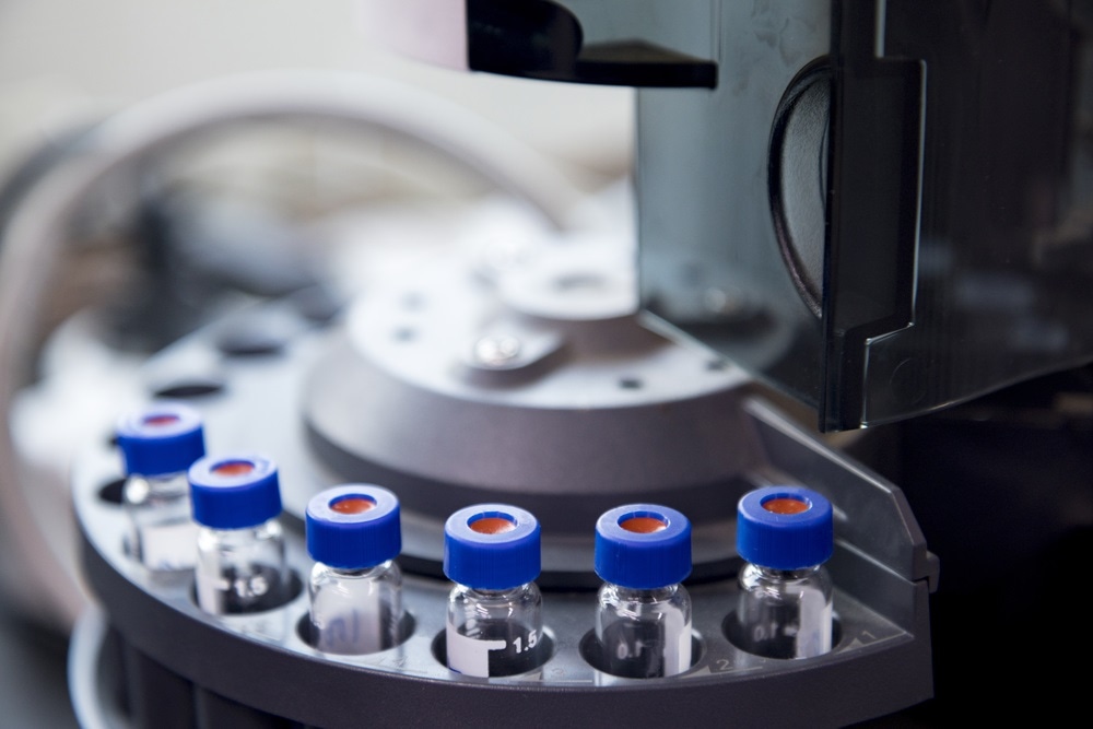 Image Credits: kdsoo322 / Shutterstock.com
How crucial are studies into the proteome to advance our understanding of human diseases?
It is incredibly important to study the proteome to understand human diseases. There are two different ways in which looking at the proteome can be advantageous: the first is looking at diagnostics, and the second is understanding treatment options.
For example, if you have a patient who is developing a disease, often that disease will be triggered by the expression of proteins that either would not be present in the healthy state or potentially would be present in different amounts in a healthy state versus a diseased state.
By studying the proteome and looking at what proteins are present and the quantity present, we can figure out, for example, if a patient has a particular disease. That is the first huge application.
The second application in which proteome studies can be very useful is looking at treatment options. Again, for example, if you have different drugs that can be used to treat a disease, you can use the proteins as a readout to see whether or not your drug is working, and if a different drug needs to be used for that patient. We think of proteomics as a very good readout on the health and viability of human cells.
What are some of the advances that have been made in mass spectrometry methods?
Over the last five to ten years, we have seen enormous advances in the area of mass spectrometry instrumentation as evidenced by the growing popularity of the technique. If you attend the American Society for Mass Spectrometry Meeting, it just gets bigger and bigger every year. This is in large measure because anything can be studied by looking at its molecular weight.
Some of the really exciting advances that have occurred in the last few years are the introduction of techniques like ion mobility, which allows you to discriminate between molecules that have the same molecular weight but different shapes.
With mass spectrometry, you are usually studying things and looking at differences in weight. When we add ion mobility at the front end of a mass spectrometer, we can look at things that have the same weight but different shapes. So, we are reaching an exciting point where we can look at almost anything.
In my area of research, I do a lot of proteomics and a lot of imaging mass spectrometry. There have been many developments in the world of imaging as well that allow us to look at, with really exquisite spatial resolution, just a few cells at a time in a spatially discrete manner by mass spectrometry. I am also very excited by those advances.
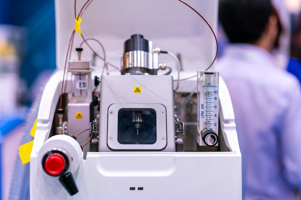 Image Credits: Surasak_Photo / Shutterstock.com
How does this type of innovation enable research that goes beyond a standard analysis?
With these new types of methods, we can start asking questions that we could not get at before. To go back to the question of ion mobility, you are using ion mobility in different types of research. Let us say, for example, you had two molecules that were exactly the same mass.
A lot of smaller molecules, like lipids, will often have the same mass, so we could not discriminate between these various species previously. But now, with these new advances, we can start figuring out these subtle differences.
Human cells have been doing this for thousands of years; they are really good at discriminating and using these different molecules. We are now just catching up with the instrumentation, and being able to discriminate and use these is very advantageous.
Why do you choose to focus on this research and what are the benefits of the research?
I was trained as an analytical chemist. I worked in an analytical chemistry group as a graduate student, and I love how fundamental and widely applicable analytical chemistry. At the end of the day, all science is driven by measurement. If you do not know what you have, you really cannot do research. Analytical chemistry is the area of research that makes all that possible. I love that aspect of it.
I also love method development and being able to develop these rigorous robust methods. I like doing analytical chemistry in the area of biomedical research because I fundamentally want to do research that will help people. That is the overarching goal of research in general.
My family has a history of certain types of cancers and so we have focused all the efforts of my research group in applying analytical chemistry to cancer biology and trying to use these measurement strategies to explore compelling questions in cancer biology.
What is in store for the future of your research in cancer biology?
My research group does a lot of work using different types of cancer model systems. More specifically, the work over the last 10 years has been using immortalized cell lines to form three-dimensional cell cultures, which are mimics of human tumors.
We have developed a suite of mass spectrometric methods that are very robust and rigorous, and we spent 10 years developing these methods so that we could then translate them from the cell culture models into actual patient tissues. We are now making this transition and moving our analytical methods from cell lines into patient-derived organoids.
These are samples that are grown from patient biopsies and we can use our mass spectrometric methods, for example, to figure out whether or not a patient will respond to a specific drug. We can take one of these biopsies, culture it, treat it with the drug, and use mass spectrometry to see whether or not the patient can metabolize that drug. Having that information can help guide clinical decisions.
.jpg&ts=20200618120202&ri=1000) Image Credits: CI Photos / Shutterstock.com
Image Credits: CI Photos / Shutterstock.comWhat other research focuses are there at Pittcon?
I have given a talk on using the proteome to look at ways to increase chemotherapeutic efficacy. My lab has been investigating different nutritional modifications to investigate the question of whether or not we can make chemotherapy work better. We have some very promising preliminary data indicating that eating less can increase chemotherapeutic efficacy in colon cancer cells. This is based on both phenotypic and proteomic data.
I’ve also given a talk on my research group’s imaging mass spectrometry work, using our three-dimensional spheroid models and organoids to look at both pharmacokinetics and pharmacodynamics of different compounds that are available for colorectal cancer patients.
With both of these talks, we were looking to apply mass spectrometry to what I consider really important problems in colorectal cancer research, and hopefully come up with better options for patients.
What do you hope to gain from Pittcon with these research presentations?
It is always wonderful to come here because you get to see all your friends and colleagues as well as meet new people. During one of my talks, I met a bunch of people I had never met before and got to hear about their research and some of the ideas and concepts that were introduced in the symposium were things I had not thought of.
That changes my perspective a little bit on the research and sometimes it forces you to focus on other aspects or hone your hypotheses a little bit. I enjoy being introduced to these new ideas.
Also, if you get really good questions after you give a talk it can sometimes be hard to answer them, but it also can make your research that much better if it forces you to think about why you are doing what you are doing. I find Pittcon to be a very rich scientific environment and there are a lot of good discussions going on here.
Why do you think Pittcon benefits the analytical science community?
I have been coming to Pittcon since I was a grad student; I think this is my 15th year here, perhaps. It is always so important to come and to interact with your friends and colleagues to find out what is going on in the field and to get a sense of where the community is moving.
It has also been neat to see changes in the focus of analytical chemistry over the years and to see different topics becoming emphasized and more important.
About Amanda Hummon – Ohio State University
As a member of the Molecular Carcinogenesis and Chemoprevention at the OSUCCC – James, Amanda’s research interests lie at the intersection of analytical chemistry and chemical biology, with a focus on cancer biology.
The lab develops analytical methods to evaluate the proteome, phosphoproteome and metabolome in cancer cells while exploring the deregulation in cancer-associated signal transduction pathways. We use global mass spectrometric profiling to quantitatively assess the altered abundance of biomolecules in cancer cells, particularly in colon cancer. We have used this approach to examine patient samples, mouse xenografts and immortalized cell lines.
Amanda’s lab also develops imaging mass spectrometry methods to track the penetration and metabolism of chemotherapies in tumors and three-dimensional cell cultures.
Comments are closed.